瀏覽量: 119
- 產(chǎn)品名稱: Anti Active Rac Mouse Monoclonal Antibody
- 產(chǎn)品貨號(hào): CST26903
- 貨期: 現(xiàn)貨
- 價(jià)格與訂購: 4800
- 數(shù)量:
庫存: 5
- 規(guī)格: 100μg
- 產(chǎn)品信息
- 如何訂購
Catalog Number
CST26903
Gene Symbol
RAC1
iconicon
Description
Anti Active Rac1-GTP Mouse Monoclonal Antibody
Background
Small GTPases are a super-family of cellular signaling regulators. Rac belongs to the Rho sub-family of GTPases that regulate cell motility, cell division, and gene transcription. GTP binding increases the activity of Rac, and the hydrolysis of GTP to GDP renders it inactive. GTP hydrolysis is aided by GTPase activating proteins (GAPs), while exchange of GDP for GTP is facilitated by guanine nucleotide exchange factors (GEFs).
Immunogen
Recombinant full length protein of active Rac1
Applications
IP, IHC
Recommended Dilutions
1 μg for 1~2 mg total cellular proteins for IP;
1:100 dilution for IHC
Concentration
1 mg/ml
Host
mouse
icon
Format
Liquid
Clonality
Monoclonal
Isotype
IgM
Purity
Purified from ascites
Preservative
no
Constituents
PBS (without Mg2+ and Ca2+), pH 7.4, 150 mM NaCl, 50% glycerol
Species Reactivity: Anti-active Rac1 antibody recognizes active Rac1 from vertebrates.
Storage Conditions: Store at -20°C. Avoid freeze / thaw cycles
Species Reactivity
Anti-active Rac1 antibody recognizes active Rac1 from vertebrates.
Immunoprecipitation/Western Blot

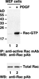
Rac activation assay. MEF cells were treated with (lane 2) or without (lane 1) PDGF. Cell lysates were incubated with an anti active Rac monoclonal antibody (Cat # 26903) (top panel). Th precipitated active Rac was immunoblotted with an anti Rac rabbit polyclonal antibody (Cat # 21003). The bottom panel shows the Western blot with anti Rac of the cell lysates used (5% of that used in the top panel).
icon
Storage Conditions
Store at -20°C. Avoid freeze / thaw cycles
iconicon
Immunofluorescence:

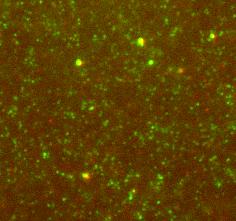
Immunofluorescence for the active Rac using Anti Active Rac1 GTP Mouse Monoclonal Antibody [26903] shows Rac GTP immunolabeling (green) in combination with cofilin (red) on brain tissue sections.
The tissue sections were fixed with -20 oC methanol or 4% paraformaldehyde (fixation time 1hr) and stained with antibody at 1:1000 in 0.1M Phosphate buffer with 0.3% Triton X, and 4% BSA for 24h at room temperature. Secondary antibodies were anti mouse AlexaFluor488 and anti rabbit AlexaFluor594 at 1:1000.
icon Note
For Research Use Only.

 地 址:
地 址: 產(chǎn)品銷售:
 E - mail :
 郵 編:
郵 編:
 Amily
Amily


